The Levi laboratory is proud to announce a publication in Wound Repair and Regeneration entitled “Short Wave Infrared Light Imaging Measures Tissue Moisture and Distinguishes Superficial from Deep Burns.” We are excited to share a novel imaging modality that may be the future of care for patients with burn injuries. In the operating room, it can be difficult to distinguish between the almost dead and the completely dead tissues.
Read more about how this new moisture detecting technology may provide a long awaited tool for the burn surgeon. This work showcases the interdisciplinary collaboration central to the University of Michigan, driven by Dr. Sergey Mironov (Berenfeld Lab, Department of Engineering), Charles Hwang (Levi Lab, Department of Surgery), and Dr. Jean Nemzek (Unit for Laboratory Animal Medicine). This work may serve as the proof of concept that paves the way for exciting healthcare innovations and technologies for patient care. Special thank you’s to the rest of the Levi laboratory team.
PMID: 31675450 (Read here!)
Abstract
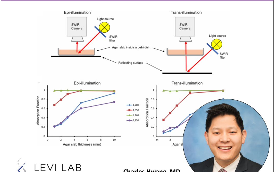
Existing clinical approaches and tools to measure burn tissue destruction are limited resulting in misdiagnosis of injury depth in over 40% of cases. Thus, our objective in this study was to characterize the ability of short-wave infrared (SWIR) imaging to detect moisture levels as a surrogate for tissue viability with resolution to differentiate between burns of various depths. To accomplish our aim, we constructed an imaging system consisting of a broad-band Tungsten light source; 1,200-, 1,650-, 1,940-, and 2,250-nm wavelength filters; and a specialized SWIR camera. We initially used agar slabs to provide a baseline spectrum for SWIR light imaging and demonstrated the differential absorbance at the multiple wavelengths, with 1,940 nm being the highest absorbed wavelength. These spectral bands were then demonstrated to detect levels of moisture in inorganic and in vivo mice models. The multiwavelength SWIR imaging approach was used to diagnose depth of burns using an in vivo porcine burn model. Healthy and injured skin regions were imaged 72 hours after short (20 seconds) and long (60 seconds) burn application, and biopsies were extracted from those regions for histologic analysis. Burn depth analysis based on collagen coagulation histology confirmed the formation of superficial and deep burns. SWIR multispectral reflectance imaging showed enhanced intensity levels in long burned regions, which correlated with histology and distinguished between superficial and deep burns. This SWIR imaging method represents a novel, real-time method to objectively distinguishing superficial from deep burns.